lipid rich adrenal adenoma
Lipid-rich Adenoma Adrenal Gland. 53 rows if an adrenal mass measures 10 hu or less on unenhanced ct it is probably a lipid-rich adenoma.

Adrenal Adenoma Radiology Case Radiopaedia Org
The present study was undertaken to evaluate the hypothesis that lipid-rich adrenal incidentalomas a hallmark of benign adrenal adenomas may not show excess growth andor develop excess hormonal secretion during short-term follow-up and that it might be possible to re-evaluate them after 5-year follow-up instead of at 1 to 2 years intervals.

. These values were consistent with lipid-rich adenoma. An adrenal adenoma is a benign or non-cancerous tumor of the adrenal gland located just above the kidney. Approximately 70 of adrenal adenomas have a high intracytoplasmic lipid content and therefore have a readily distinguishable phenotype as a lowattenuation. Most benign adrenal tumors cause no symptoms and dont need treatment.
A lipid-rich adrenal tumor presenting increased FDG uptake compared with that of the liver is likely to be a hormone-secreting adenoma. Some such adenomas called non-functioning adrenal adenomas do not secrete hormones but others do. However some can become functioning or active and secrete excess. For lipid-rich adenomas of the adrenal glands measuring under 4 cm in a patient with no underlying malignancy no follow up imaging is required 1.
They are common and they usually. The premise with this technique is that most adenomas contain lipid and more than 80 are of the lipid-rich variety 11. Most adrenal gland adenomas dont cause any problems -- they just take up space. 14 the mean hounsfield unit for adrenal carcinoma metastasis and pheochromocytoma is significantly.
Thirty-five surgically resected adrenal adenomas were used. Those that develop in the medulla are also called pheochromocytomas fee-o-kroe-moe-sy-TOE-muhs. The front line treatment for Adrenal Adenoma is surgery. Up to 10 cash back The T2W properties of a lipid-rich adenoma may be useful to establish diagnosis when differentiating lipid-rich adenoma from other adrenal masses which may demonstrate microscopic fat on chemical-shift MRI or when chemical-shift MRI is degraded by artifact or limited by through-plane resolution in small adrenal nodules.
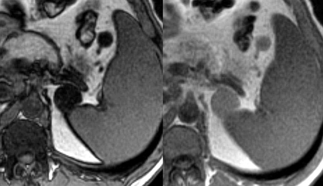
Lipid-rich adenomas lose signal on the chemical-shift or out-of-phase opposed-phase images while lipid-poor lesions will not lose signal. In general malignant adrenal lesions do not contain lipid. The lower the Hounsfield Units lipid-rich. Adrenal adenomas are one of several types of nodules that develop on the adrenal glands.
Im writing on behalf of my mother XXXX who is 72 years old. My mother recently went to a doctor and here is the doctors report below that she received. D E Axial T1 and T2-weighted MR images showed a well-defined left adrenal mass displaying isointense signal relative to spleen on T1 and T2WIs. A high-quality CT scan using contrast is the most important x-ray or scan.
Adrenal Adenoma is a pathological condition of the adrenal glands in which there is development of benign tumors in the adrenal glands. A density equal to or below 10 HU is considered diagnostic of a lipid-rich adenomas. I am asking what do you recommendprognosis at this point. On the basis of the evaluation of qualitative chemical-shift CS signal intensity SI loss adrenal adenomas were respectively divided in Group 1A n 25 as lipid-rich and Group 1B n 10.
But some of them are functioning tumors -- that means they make the same hormones as. If it does not lose SI all one can state is that the lesion does not contain lipid and is therefore not a lipid-rich adenoma. To evaluate the relationship between lipid-rich cells of the adrenal adenoma and precontrast computed tomographic CT attenuation numbers in three clinical groups. The clinical diagnoses of the patients included 13 cases of primary aldosteronism 15.
Frequently adenomas contain abundant intracytoplasmic lipid and thus approximately 70 of adrenal adenomas are lipid-rich and are readily diagnosed because these lesions measure 10 HU or greater on unenhanced CT 2. On MR lipid-rich adrenal adenomas may demonstrate out-of-phase signal dropout which again demonstrates that the lesion is a benign adenoma despite FDG avidity Fig. Adrenal metastases should not have Hounsfield units of less than 10 on unenhanced CT. Lipid-rich adenoma 70 of adenomas contain high intracellular fat and will be of low attenuation on unenhanced CT 45.
The surgical procedure done for removal of Adrenal Adenoma is called as adrenalectomy. The marked reduction in signal intensity between the in-phase and out of phase T1-weighted images indicates fatty content and therefore a lipid-rich adenoma. 1516 Delayed contrastenhanced phase CT scans are performed to further distinguish between those 30 of benign tumours with low lipid content and malignant. F G coronal gradient-echo in-phase and out-of-phase MR images showed significant visual signal loss between in-phase and out-of-phase images.
Benign adrenal tumors that develop in the cortex are also called adrenal adenomas. The absolute or relative percentage washout of contrast material on delayed contrast-enhanced CT is a highly specific test for the differentiation of lipid-poor and lipid-rich adrenal adenomas from adrenal nonadenomas. Know the causes symptoms treatment and prognosis of adrenal adenoma. Chemical-shift MRI is now the most commonly used imaging technique to distinguish between adenomas and metastases.
But sometimes these tumors secrete high levels of certain hormones that can cause. They are often found incidentally during imaging studies of the abdomen in which case they are referred to as adrenal incidentalomas. Fatty are the more likely it is that the tumor is not a cancer but rather the more common adrenocortical adenoma. Differentiation of adrenal adenomas lipid rich and lipid poor from nonadenomas by use of washout characteristics on delayed enhanced CT.
What do these following CT scan findings indicate. Therefore additional endocrinological investigations are strongly recommended when an FDG-avid lipid. Adrenal metastases should not demonstrate out-of-phase signal dropout on MR. The majority of adrenal adenomas are nonfunctioning which means they do not produce hormones and usually do not cause any symptoms.
Depending upon the type of hormone secreted by the adenoma the tumor can cause different medical problems in the patient. Thus if a lesion loses SI it is a lipid-rich adenoma Fig.

X Rays Ct Scans Mri And Other Tests For Adrenal Glands

Hca Hospital Adrenal Gland Tumor Screening Program

Chemical Shift Mr And Precontrast Ct Scans Of Right Lipid Rich Adrenal Download Scientific Diagram

Posting Komentar untuk "lipid rich adrenal adenoma"